In modern implant dentistry, predictable outcomes rely heavily on the quality and quantity of the underlying bone. Without sufficient bone volume, dental implants may fail to achieve long-term stability or optimal aesthetics. This is where bone grafting becomes indispensable. By restoring or augmenting the alveolar bone, clinicians can create a solid foundation for implants, enhance osseointegration, and improve overall treatment predictability.
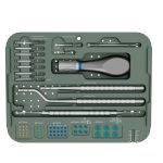
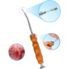
Bone grafting is particularly important in cases of tooth loss, periodontal disease, trauma, or anatomical limitations. Additionally, advances in surgical tools, such as the DentalMaster Bone Screw Kit and Auto Bone Tack Kit, allow for precise fixation of grafts, reduced surgical time, and improved clinical outcomes.
1. In-Depth Understanding of Bone Grafting in Dental Implantology
Bone grafting, as a critical adjunctive technique in the field of dental implantology, refers to the reconstruction of alveolar ridge defects caused by periodontal disease, trauma, or congenital deficiencies through the implantation of autogenous bone, allogeneic bone, or synthetic bone substitute materials. The core purpose of this process is to provide sufficient bone volume and ideal bone density for subsequent implant placement, ensuring that the implant can achieve stable mechanical support and long-term stability.
From a biological perspective, successful bone grafting not only fills the defect area but, more importantly, activates bone regeneration signaling pathways, promoting the proliferation and differentiation of bone cells (such as osteoblasts). These bone cells gradually grow and adhere to the implant surface, ultimately forming a biomechanically robust bone–implant interface, known as osseointegration. This process is crucial for the long-term success of the implant, ensuring its durable stability under the complex mechanical environment of the oral cavity.
In clinical practice, experienced clinicians comprehensively evaluate the specific size of the defect, its anatomical location, as well as the patient’s overall health and local bone conditions. This assessment allows them to precisely select the most suitable type of graft material (such as particulate, block, or membrane forms) and grafting technique (such as guided bone regeneration or sinus lift procedures) in order to achieve the optimal therapeutic outcome.
2. Clinical Indications for Bone Grafting
2.1 Tooth Loss
After tooth extraction, alveolar bone naturally resorbs, sometimes leading to insufficient width or height for implant placement. Socket preservation techniques, often using graft material and a fixation system like the Auto Bone Tack Kit, can prevent significant bone loss.
2.2 Periodontal Disease
Chronic periodontal infections cause localized bone defects around teeth. Bone grafting can restore lost bone, providing a stable site for implant placement while improving long-term periodontal health.
2.3 Trauma or Infection
Fractures, tooth avulsion, or localized infections can result in bone deficiencies. Bone grafting allows clinicians to rebuild the ridge architecture and create a suitable environment for implants.
2.4 Sinus Pneumatization and Maxillary Deficiency
In the posterior maxilla, sinus expansion can reduce available bone height. Sinus lift procedures with grafting are commonly performed to increase vertical bone volume, enabling implant placement in otherwise compromised areas.
3. Types of Bone Graft Materials and Techniques
3.1 Autogenous Grafts
Harvested from the patient’s own body, these grafts offer the highest osteogenic potential. Common donor sites include the chin, mandibular ramus, and iliac crest.
3.2 Allografts and Xenografts
Processed human or animal bone substitutes provide structural support and volume maintenance. They are widely used when autogenous bone is insufficient or to reduce surgical morbidity.
3.3 Alloplastic Materials
Synthetic grafts, such as hydroxyapatite or beta-tricalcium phosphate, offer predictable resorption rates and biocompatibility.
3.4 Membranes and Fixation Systems
To ensure graft stability and prevent soft tissue ingrowth, clinicians use resorbable or non-resorbable membranes. Tools like the DentalMaster Bone Screw Kit or Auto Bone Tack Kit secure both block and particulate grafts effectively, enhancing integration and minimizing movement.
4. Surgical Techniques in Bone Augmentation
4.1 Socket Preservation
Performed immediately after tooth extraction, socket grafting minimizes bone resorption. The graft is placed in the extraction socket and stabilized, often with micro-tacks or screws for optimal results.
4.2 Block Grafting
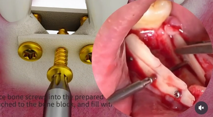
Used for significant ridge deficiencies, block grafts are harvested, shaped, and fixed onto the deficient ridge. The Bone Screw Kit simplifies the stabilization process, ensuring precise fixation.
4.3 Lateral Ridge Augmentation
This technique increases ridge width to accommodate implants. Particulate grafts are secured with tacks or screws and covered with a membrane for guided bone regeneration.
4.4 Sinus Lift Procedures
For posterior maxillary implants, graft material is placed under the elevated sinus membrane. Fixation tools ensure graft stability while minimizing membrane perforation risk.
5. What Happens Before, During, and After Bone Grafting
5.1 Preoperative Assessment
- CBCT scans to evaluate bone volume and quality
- Identification of potential anatomical challenges
- Discussion of graft material and fixation system selection
5.2 Surgical Procedure
- Local anesthesia or conscious sedation
- Flap elevation and defect exposure
- Placement of graft material, secured with Bone Screw Kit or Auto Bone Tack Kit
- Membrane coverage for guided regeneration
5.3 Postoperative Care
- Monitoring for infection or swelling
- Soft diet for initial healing period
- Prescribed analgesics and antibiotics as needed
6. Post-Surgical Considerations and Maintenance
- Healing typically spans 3–6 months before implant placement
- Signs of successful integration include bone density increase and stability of graft
- Follow-up imaging and clinical evaluation are essential
- Early intervention in case of exposure or infection improves outcomes
7. Long-Term Benefits of Dental Implants
First, through scientifically precise implant placement techniques and individualized design, dental implants can significantly enhance primary stability, providing a solid foundation for subsequent osseointegration. Their excellent support capability effectively distributes occlusal forces, protecting surrounding bone tissue and the health of adjacent teeth.
Second, in the anterior aesthetic zone, dental implants, through precise three-dimensional positioning and customized restorative planning, can achieve a highly natural coordination of gingival contours, tooth morphology, and color, resulting in predictable and long-lasting aesthetic outcomes that meet patients’ personalized smile requirements.
Moreover, owing to their favorable biocompatibility and mechanical properties, dental implants can maintain long-term masticatory function, reduce discomfort such as food impaction, and significantly improve patients’ oral function and quality of life, thereby enhancing overall satisfaction.
Finally, by optimizing the bone–implant interface design and stress distribution, implants can minimize the risk of bone resorption, reduce the likelihood of future implant loosening or peri-implantitis, and provide patients with long-term stable oral health.
8. Future Trends in Bone Augmentation
With the widespread application of innovative tools such as the DentalMaster Bone Screw Kit and Auto Bone Tack Kit, clinicians now have more efficient solutions for implant surgery. These advanced instruments streamline surgical procedures, significantly shorten operative time, and reduce patient discomfort from prolonged mouth opening.
At the same time, their precise bone fixation design ensures that bone augmentation materials remain stable, creating an ideal microenvironment for bone tissue regeneration. Furthermore, standardized operational protocols contribute to predictable healing outcomes and lower the risk of postoperative complications.
The design philosophy of these tools closely aligns with the core requirements of modern implant surgery: efficiency, precision, and reliability, providing strong technical support for clinical practice.
9. Conclusion
Bone grafting, as a core method for addressing insufficient bone volume, is a critical step in achieving long-term success with dental implants. In complex clinical situations, such as inadequate alveolar ridge height or width, scientifically standardized bone grafting techniques can reconstruct suitable bone conditions, providing a stable biological foundation for implants.
In clinical practice, by developing personalized treatment plans, precisely selecting biocompatible graft materials, and incorporating innovative fixation devices such as the Bone Screw Kit and Auto Bone Tack Kit, clinicians can effectively restore the three-dimensional structure of the bone, significantly enhancing both primary stability and long-term retention of implants.
This integrated approach not only ensures predictable outcomes in terms of function and aesthetics but also improves treatment efficiency through minimally invasive and precise procedures. For dental professionals, systematically integrating such advanced technological systems into clinical practice can optimize surgical outcomes, reduce complication risks, and enhance patient confidence and long-term satisfaction through improved treatment experience and restorative quality.