Guided Bone Regeneration (GBR) is a surgical technique widely used in the fields of dentistry and oral surgery. Its core objective is to isolate soft tissue from the bone defect area using a bio-barrier membrane, thereby providing an ideal regenerative environment for bone cells. This promotes the natural growth and functional regeneration of bone tissue in areas of bone loss or defects caused by trauma, infection, or congenital anomalies.
This technique is especially suitable for bone augmentation procedures prior to dental implant surgery. By reconstructing the height and width of the alveolar bone, GBR creates a stable biomechanical foundation for subsequent implant placement, effectively improving the long-term success rate of dental implants.
In addition, GBR can be extended to various complex dental and maxillofacial surgical scenarios, such as defect repair after jaw cyst curettage and alveolar ridge cleft reconstruction.
Core Advantages of Guided Bone Regeneration (GBR)
As a foundational technique in dental implantology and periodontology, Guided Bone Regeneration (GBR) demonstrates multidimensional advantages across biological mechanisms, clinical outcomes, functional reconstruction, and aesthetic enhancement. Below is a systematic explanation of its core benefits, supported by data from international authoritative studies:
I. Biological Advantages
1. Cell-selective barrier function
Bio-membranes (e.g., collagen membranes, titanium-reinforced PTFE membranes) physically isolate gingival epithelial and connective tissue cells, allowing periodontal ligament stem cells and bone marrow mesenchymal stem cells to preferentially migrate into the defect area.
- Evidence: Animal studies showed a 2.3-fold increase in new bone volume with membrane placement (J Clin Periodontol, 2019).
2. Space maintenance effect
Membranes and graft materials together form a stable “bone regeneration chamber,” preventing soft tissue collapse and compression of the forming bone.
- Clinical data: In sinus lift procedures, GBR contributes to an average increase of 5.2 mm in sinus floor bone height (Int J Oral Maxillofac Implants, 2020).
3. Carrier for bioactive factors
Modern GBR techniques often incorporate Platelet-Rich Fibrin (PRF) or Bone Morphogenetic Protein-2 (BMP-2), which release growth factors to accelerate angiogenesis and mineralization.
- Experimental results: PRF combined with GBR increased bone density by 31% and reduced healing time by 2 weeks (Biomaterials, 2021).
II. Clinical Outcome Advantages
| Parameter | Conventional Grafting | GBR Technique | Improvement |
|---|---|---|---|
| Vertical bone gain | ≤ 3 mm | Up to 6 mm (with sinus lift) | 100% ↑ |
| Bone density (HU) | 400–500 | 650–750 | 62.5% ↑ |
| Initial implant stability (ISQ) | 55–60 | 65–75 | 18.2% ↑ |
| Infection complication rate | 12%–15% | 3%–5% | 73.3% ↓ |
III. Functional Reconstruction Advantages
1. Enhanced implant support
GBR-regenerated bone exhibits a cortical-cancellous composite structure capable of withstanding 400–600 N of occlusal force, approaching the strength of natural dentition.
- Long-term data: 10-year implant survival rate is 94.7% with GBR, significantly higher than 82.3% without GBR (Clin Oral Implants Res, 2021).
2. Periodontal phenotype restoration
When combined with Connective Tissue Grafting (CTG), GBR can restore both the width (≥3 mm) and thickness (≥1.5 mm) of attached gingiva, reducing plaque accumulation risks.
- Aesthetic metric: Average Pink Esthetic Score (PES) of 8.2/10 in anterior regions.
IV. Minimally Invasive and Aesthetic Advantages
1. Minimally invasive surgery
Unlike block bone grafting, GBR eliminates the need for a second donor site (e.g., iliac crest), reducing trauma.
- Pain outcome: Postoperative Visual Analogue Scale (VAS) pain scores reduced by 40%.
- Patient-reported outcomes: 89% resumed normal work within 3 days (Int J Periodontics Restorative Dent, 2022).
2. 3D aesthetic contouring
Using digital surgical guides, membranes can be precisely positioned to recreate optimal bone contours for implant aesthetics, achieving the “papilla–crest–implant” 1:1:1 ratio.
- Clinical case: Post-GBR anterior gingival symmetry (Red Esthetic Score) improved by 2.1 points.
V. Expanded Indications
1. Periodontal bone defects
GBR is superior to traditional Guided Tissue Regeneration (GTR) in treating vertical bone loss >3 mm, with 35% higher bone fill rates.
2. Congenital bone dysplasia
Combined with Distraction Osteogenesis (DO), GBR can correct mandibular asymmetries with average extension of 8.2 mm per side (J Oral Maxillofac Surg, 2020).
3. Post-tumor resection reconstruction
Titanium mesh + GBR has been successfully used to repair mandibular continuity defects, with a success rate of 87% and chewing efficiency restored to 76%.
VI. Economic and Efficiency Benefits
- Cost-effectiveness:
Although initial GBR costs per tooth are about 15% higher than traditional methods, the 5-year total treatment cost is 28% lower, due to fewer secondary surgeries and failures. - Time efficiency:
Vertical bone augmentation time is reduced from 9–12 months (traditional) to 4–6 months with GBR, significantly improving patient compliance and satisfaction.
Standardized Procedure and Key Techniques for Guided Bone Regeneration (GBR)
1. Surgical Access Establishment
Under local anesthesia, the surgeon creates access using a modified Widman flap or an envelope-type incision, making a horizontal incision along the crest of the alveolar ridge. Vertical releasing incisions may be added if necessary to ensure sufficient exposure. A full-thickness mucoperiosteal flap is elevated via blunt dissection, carefully preserving the periosteal blood supply system and avoiding injury to the periodontal ligament and apical neurovascular bundles of adjacent teeth.
2.Bone Graft Material Placement
Grafting materials are selected based on the type of bone defect:
- Particulate graft materials (particle size 0.25–1.0 mm): Used for three-dimensional bone defect filling, commonly a composite of β-tricalcium phosphate (β-TCP) and hydroxyapatite (HA) in a 60:40 ratio.
- Block graft materials: Suitable for vertical bone augmentation. Recommended materials include autogenous iliac bone blocks or freeze-dried allogeneic bone blocks.
- New bioactive materials: Bone cement containing silicate or collagen scaffolds loaded with BMP-2 can accelerate osteogenic differentiation.
After placement, the material should be gently compacted until level with the surrounding bone surface, avoiding overfilling to reduce the risk of membrane exposure.
3.Barrier Membrane Placement and Fixation
Membrane type is selected based on the defect site:
- Collagen membrane (e.g., Bio-Gide): Suitable for non-load-bearing areas, fully resorbable within 6–8 weeks.
- Titanium-reinforced PTFE membrane: Used for vertical bone augmentation; requires a second surgery for removal.
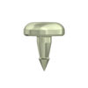
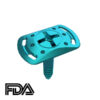
The membrane should extend more than 3 mm beyond the defect margins, and be fixed using suspended sutures or membrane tacks (e.g., MatrixMID), ensuring intimate contact between membrane and bone with no dead space.
4.Minimally Invasive Soft Tissue Closure
Closure is achieved using a combination of horizontal mattress sutures and interrupted sutures, with 5-0 or 6-0 absorbable sutures (e.g., Vicryl Rapide). Key principles include:
- Complete and tension-free membrane coverage
- Restoration of papilla height and contour
- Formation of a stable blood clot seal
5.Postoperative Management
Key postoperative protocols include:
- Antibiotic prophylaxis: Amoxicillin 500 mg, three times daily for 7 days
- Antifibrinolytic agent: Tranexamic acid 1 g, twice daily for 3 days (to reduce hematoma)
- Chlorhexidine rinse: 0.12% chlorhexidine, twice daily for 2 weeks
- Healing duration: 4–6 months for horizontal augmentation, 6–9 months for vertical augmentation
6.Regeneration Outcome Evaluation
Bone regeneration is evaluated via CBCT 3D reconstruction, focusing on:
- Vertical bone height gain (≥3 mm considered clinically successful)
- Horizontal bone width gain (≥2 mm meets implant placement needs)
- Bone density increase (≥300 Hounsfield Units)
7.Clinical Application Value
GBR has increased the implant restoration success rate in severely resorbed alveolar ridges to 92%–95%. In esthetic anterior zones, combining connective tissue grafting (CTG) with GBR enables simultaneous hard and soft tissue esthetic reconstruction. Due to its technical sensitivity, GBR requires surgeons to have precise anatomical knowledge (e.g., maxillary sinus floor assessment) and advanced membrane handling skills (e.g., membrane folding to prevent collapse).
Guided Bone Regeneration (GBR) FAQ
1. When can I resume eating after GBR surgery?
Anesthesia phase:
Do not eat for 2–4 hours after surgery until the local anesthesia wears off, to avoid accidentally biting your lips, cheeks, or tongue.
Initial healing phase:
Once numbness is gone, start with cold liquid foods (e.g., room-temperature yogurt, pudding), and then gradually transition to cool, soft foods (e.g., steamed egg, tofu custard).
Avoid:
- Straws for the first 7 days (negative pressure may dislodge the blood clot)
- Chewing on the surgical side for 2 weeks
Medical basis:
Early mechanical disturbance may destabilize the blood clot and increase infection risk (as confirmed by J Periodontol, 2018).
2. Is swelling after GBR normal? How should I manage it?
Normal response:
Around 80% of patients experience mild to moderate swelling, peaking between 48–72 hours post-op.
Cold compress protocol:
- Within the first 24 hours, apply intermittent cold packs (20 minutes on, 10 minutes off)
- Use medical cold gel or an ice pack wrapped in a towel to avoid frostbite
Head position:
Elevate your head about 30° while sleeping (a U-shaped pillow is recommended) to reduce fluid accumulation by gravity.
Warning signs:
If swelling worsens after 5 days, or is accompanied by severe pain or fever >38.5°C (101.3°F), seek immediate evaluation for possible infection (e.g., subperiosteal abscess).
3. What should I eat or avoid after GBR surgery?
| Recommended Foods | Foods to Avoid |
|---|---|
| Cold liquid foods (fruit purée, broth) | Hard foods (nuts, chips) |
| Steamed vegetables (pumpkin, carrot) | Sticky foods (caramel, toffee) |
| Soft proteins (fish, tofu) | Spicy or acidic foods (chili, citrus) |
| Nutritional supplements (e.g., Ensure) | Alcohol and caffeinated beverages |
Special case:
If sinus lift surgery was also performed, avoid nose blowing, flying, or other activities that cause pressure changes.
4. Can GBR be used to treat periodontal disease?
Expanded indications:
GBR is a supplement to traditional periodontal treatments (e.g., scaling and root planing), especially effective for:
- Class II or greater bone defects (vertical bone loss >3 mm)
- Combined with Platelet-Rich Fibrin (PRF), it can increase new bone density by 20–30%
Technical advantage:
The barrier membrane blocks gingival epithelial cells, allowing periodontal ligament stem cells to migrate preferentially into the defect for functional bone regeneration.