
Dental bone grafting, as a crucial and widely applied technique in the field of oral surgery, plays an irreplaceable role in repairing alveolar bone defects and reconstructing jawbone structures. In clinical dental practice, due to situations such as long-term tooth loss without timely restoration, severe periodontal disease erosion, or trauma from external impacts, the alveolar bone often undergoes varying degrees of atrophy and resorption, leading to insufficient bone volume. This not only affects the normal function of the teeth but also negatively impacts the patient’s facial aesthetics.
Dental bone grafting technology emerged precisely to address these issues. Its core purpose is to provide stable bone support for subsequent dental implant procedures by grafting appropriate bone materials, ensuring that the implants can firmly integrate with the bone tissue and achieve long-term functional stability. At the same time, for patients with jawbone deformities caused by bone defects, this technique can effectively restore the normal shape and structure of the jawbone and help regain oral physiological functions.
This article will integrate authoritative medical literature and extensive clinical experience to comprehensively and systematically outline the indications for dental bone grafting, clarifying under what circumstances this technique is appropriate. It will detail the surgical procedures, allowing readers to understand every key step from preoperative preparation to intraoperative operation and postoperative management. It will deeply explore material selection, analyzing the characteristics, advantages and disadvantages, and applicable scenarios of different bone grafting materials. At the same time, it will emphasize the key points of postoperative care, including oral hygiene, dietary adjustments, and how to deal with potential complications.
The aim is to provide patients with scientific and comprehensive treatment references to help them better understand bone grafting and actively cooperate with treatment. It also seeks to offer practical clinical guidelines for oral surgeons, improve treatment quality, and promote broader application and development of bone grafting techniques in the field of dentistry.
What is dental bone grafting?
Dental bone grafting is an important technique in the field of oral surgery. It involves transplanting autogenous bone, allogeneic bone, or synthetic bone materials into areas with alveolar bone defects or abnormal jawbone structures. It is used to supplement bone volume in regions with insufficient bone caused by long-term tooth loss, periodontal disease, trauma, and other conditions. The goal is to repair the damaged alveolar bone, reconstruct the normal structure of the jawbone, provide a stable and solid bone foundation for subsequent dental implant procedures, or to improve problems such as impaired chewing function and facial aesthetics affected by bone defects, thereby helping patients restore oral health and normal physiological functions.
Indications and Applications of Dental Bone Grafting
Alveolar bone atrophy is often a major indication for dental bone grafting. Many patients experience gradual bone resorption and atrophy due to long-term tooth loss without timely restoration, as the alveolar bone lacks physiological stimulation. Severe periodontal disease can destroy the periodontal tissues and further invade the alveolar bone, leading to continuous reduction in bone height and width. Trauma may also directly cause damage or defects to the alveolar bone. Once the alveolar bone height is less than 1 cm and the width is less than 0.6 cm, it can no longer provide sufficient support and stability for dental implants. In such cases, dental bone grafting is necessary to increase bone volume and create favorable conditions for subsequent implant surgery.
Maxillary sinus lift is also a common application scenario for dental bone grafting. In the posterior maxillary region, pneumatization of the maxillary sinus often results in significantly insufficient bone volume. During dental implant procedures, if the bone volume is inadequate, the implant cannot achieve good primary stability and long-term osseointegration. Therefore, grafting materials are used to elevate the sinus floor and increase the available bone height to meet the requirements for implant placement.
Dental bone grafting is also indispensable in bone defect repair. In clinical practice, surgeries such as cyst removal or tumor curettage may inevitably leave bone cavities, and bone nonunion or bone loss may occur after fractures. These bone defects not only affect the normal structure and function of the oral cavity but may also lead to complications such as infections. Filling the bone cavities through bone grafting can promote bone tissue regeneration and repair, restoring the structural integrity of the jawbone.
In addition, dental bone grafting can be used for aesthetic and functional reconstruction. Issues such as gingival recession and root exposure not only affect dental aesthetics but may also cause discomfort like tooth sensitivity. By increasing local bone volume through bone grafting, the position and contour of the gingiva can be improved, enhancing the aesthetic outcome. At the same time, for patients requiring denture restoration, a stable bony base is key to achieving good retention and stability of the prosthesis, and bone grafting provides strong support for such restorations.
The core goal of dental bone grafting is to create a favorable microenvironment for bone regeneration at the defect site by implanting autogenous bone, allogeneic bone, or synthetic bone materials. This stimulates the activity of osteoblasts and promotes new bone formation. After a period of healing, successful osseointegration—a tight and direct bond between the grafted material and the host bone—is achieved, laying a solid foundation for subsequent treatments such as dental implants and denture restorations.
Surgical Procedure and Technical Key Points
- Preoperative Evaluation and Preparation
- Imaging Examination: Use CBCT (Cone-Beam Computed Tomography) to precisely measure the extent and density of the bone defect, as well as the surrounding anatomical structures (such as the inferior alveolar nerve and the position of the maxillary sinus).
- Systemic Health Assessment: Exclude diseases that may impair bone healing, such as uncontrolled diabetes or osteoporosis; smokers are required to quit smoking for more than 2 weeks to reduce the risk of infection.
- Anesthesia Plan: Local infiltration anesthesia or nerve block anesthesia is generally used; intravenous sedation may be combined in complex cases.
- Surgical Steps
- Incision Design: Make a trapezoidal incision along the crest of the alveolar ridge or the edge of the defect, and raise a flap to expose the bone surface. Take care to preserve the interdental papilla to maintain postoperative aesthetics.
- Recipient Site Preparation: Remove pathological tissue and smooth sharp bone edges; if sinus lifting is needed, elevate the sinus membrane using the crestal approach (osteotome technique) or lateral window technique.
- Material Grafting:
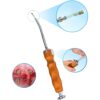
- Autogenous Bone: Harvested from the mandibular symphysis, mandibular ramus, or iliac crest; offers optimal biocompatibility but requires additional surgical incisions. Use bone scrapers to manipulate autologous bone
- Allogeneic Bone: Demineralized bone from a donor of the same species; avoids donor site complications but carries a risk of immune rejection.
- Synthetic Bone: Materials such as β-Tricalcium Phosphate (β-TCP) and Hydroxyapatite (HA), used alone or in combination, serve as osteoconductive scaffolds.
- Barrier Membrane Application: Cover the graft with a resorbable collagen membrane (e.g., Bio-Gide) to prevent soft tissue ingrowth and promote bone regeneration.
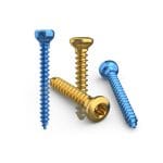
- Suturing and Fixation: Close the wound with interrupted sutures; use titanium pins if necessary to stabilize the graft material.
- Surgical Duration and Complexity
- Simple Bone Augmentation: Takes approximately 30–60 minutes, suitable for small, single-tooth defects.
- Complex Reconstruction: In cases such as full-arch alveolar bone atrophy or lateral maxillary sinus lifts, the procedure may last 2–3 hours and could involve vascularized fibula flap transplantation.
Postoperative Recovery and Complication Management
- Postoperative Care Guidelines
- Oral Hygiene: Avoid tooth brushing within the first 24 hours after surgery; instead, use chlorhexidine mouthwash. For one week, use a soft-bristled toothbrush to gently clean adjacent teeth.
- Dietary Adjustment: For the first week post-surgery, consume soft, cool or lukewarm liquid foods (such as rice soup and yogurt); avoid chewing hard foods. Refrain from eating sharp-edged foods such as nuts or crabs for one month.
- Medication Management: Take oral antibiotics (e.g., amoxicillin 500 mg, three times a day for 7 days) to prevent infection, and ibuprofen (400 mg every 6 hours) to manage pain.
- Physical Therapy: Apply ice packs within 48 hours post-surgery to reduce swelling; switch to warm compresses after 72 hours to promote blood circulation.
- Common Complications and Their Management
- Infection: Occurs in approximately 3%–5% of cases, presenting as localized redness, swelling, or exudate. Requires timely debridement and extended antibiotic therapy.
- Bone Graft Exposure: More common in patients with thin gingival biotype; can be managed by soft tissue augmentation procedures, such as free gingival grafting.
- Nerve Injury: Damage to the inferior alveolar nerve may cause numbness in the lower lip. Most cases are temporary and can be treated with oral mecobalamin (500 μg, three times a day) to promote nerve repair.
- Implant Failure: Failure of osseointegration occurs in approximately 2%–8% of cases. Requires removal of the implant and reassessment of bone conditions.
- Long-term Follow-up and Functional Maintenance
- Bone Healing Monitoring: CBCT should be reviewed 3 months after surgery to evaluate the amount of new bone formation. If bone density is less than 500 HU, implant placement should be postponed.
- Timing of Implant Placement: For autogenous bone grafts, implant surgery can usually be performed 4–6 months postoperatively; for synthetic bone grafts, the recommended interval is 6–9 months.
- Occlusal Management: After loading the implant, regular occlusal adjustments are necessary to prevent excessive loading that may lead to bone resorption.
Material Selection and Clinical Decision-Making
| Material Type | Advantages | Disadvantages | Applicable Scenarios |
|---|---|---|---|
| Autogenous Bone | High biological activity, no immune rejection | Donor site complications (pain, infection) | Complex bone defects, aesthetic zone reconstruction |
| Allogeneic Bone | Avoids donor site injury | Potential risk of disease transmission | Moderate defects, patients with limited budgets |
| Synthetic Bone (β-TCP) | Resorbable, provides osteoconductive scaffold | Lower mechanical strength | Small defects, maxillary sinus lift |
| Composite Materials | Combines advantages of autogenous and synthetic bone | Higher cost | Borderline bone volume cases |
Clinical Decision Recommendations:
- For younger patients, autogenous bone is preferred to achieve optimal osseointegration.
- For elderly or medically compromised patients, synthetic or allogeneic bone may be considered to reduce surgical trauma.
- For maxillary sinus lift procedures, a 1:1 mixture of β-TCP and autogenous bone is recommended to balance osteoconductivity and biological activity.
Future Trends in Dental Bone Grafting
The future development of dental bone grafting will revolve around four key directions: material innovation, technological intelligence, minimally invasive surgery, and personalized treatment. By integrating advanced technologies such as robotic assistance, digital navigation, and bioactive materials, maxillofacial reconstruction is moving toward greater precision, efficiency, and safety. Below is a detailed trend analysis:
1. Material Innovation: Emphasizing Both Bioactivity and Intelligence
- Superhydrophilic Materials and Osteoinductive Factors: Future materials will focus more on enhancing osseointegration efficiency. For instance, superhydrophilic titanium-zirconium implants have shown a threefold increase in wear resistance and a 20-year survival rate of 96.5%. Bioactive materials, such as Bio-Oss bone granules combined with PRF (Platelet-Rich Fibrin) growth factors, can accelerate bone regeneration and reduce the need for secondary surgeries.
- Optimization of Autogenous Tooth Bone Grafting: Due to its similar composition to bone tissue, autogenous dentin has become a new research hotspot. After demineralization to remove immunogenic components, its osteoinductive capability is significantly enhanced. In the future, standardized preparation processes may reduce composition variability and improve clinical stability.
- 3D-Printed Customized Bone Scaffolds: By integrating CT data from patients, 3D-printed scaffolds made of bioceramics or composite materials can precisely match defect morphology and carry growth factors to enable synchronized “bone-vascular” regeneration.
2. Technological Intelligence: Robotics and AI Leading Surgical Innovation
- Robotic-Assisted “One-Stop” Reconstruction: The world’s first “tooth implantation using iliac bone followed by transplantation” in 2025 marked a leap in robotic technology from simple implantation to complex maxillofacial reconstruction. In the future, robots will utilize force-motion coordinated algorithms to perform sub-millimeter osteotomies and implant placements, compressing multi-stage surgeries into a single session and shortening the treatment cycle by over 90%.
- AI Dynamic Navigation Systems: By integrating CBCT scans and intraoral 3D models, AI can generate real-time “dynamic maps” for implant paths, with accuracy controlled within 0.2 mm. This is especially beneficial in high-risk areas such as sinus lifts.
- Remote Surgical Collaboration: Technologies such as 5G + AR will enable experts to remotely control surgical robots, offering support to primary care hospitals and promoting the distribution of high-quality medical resources to lower-tier areas.
3. Minimally Invasive Surgery: From “Incision” to “Perforation”
- Minimally Invasive Incision Techniques: Implants can be placed through incisions as small as 3–4 mm. Combined with water lasers to reduce thermal damage, postoperative bleeding can be reduced by 70%, allowing most patients to resume normal eating the next day.
- Zygomatic Implant Techniques: This approach bypasses atrophic bone areas by placing implants directly into the zygomatic bone, solving the challenge of implantation in patients with severe bone loss.
- Non-Invasive Monitoring and Recovery: Wearable devices can monitor osseointegration in real time and adjust rehabilitation protocols through biofeedback, reducing the need for follow-up visits.
4. Personalized Treatment: Multidisciplinary Collaboration and Precision Matching
- Multidisciplinary Collaborative Treatment Model: Oral surgeons, prosthodontists, and orthodontists will work closely together—for example, designing integrated “bone-tooth-soft tissue” reconstruction plans for patients undergoing tumor resection.
- Genetic Testing to Guide Treatment: By analyzing patients’ bone metabolism-related genes (e.g., BMP, RUNX2), it is possible to predict osseointegration speed and customize implant surface treatments (e.g., acid-etching, sandblasting) to optimize osteogenesis.
- Aesthetic and Functional Integration: Digital Smile Design (DSD) will be integrated with bone grafting to precisely match gingival contours, crown morphology, and facial features, enhancing both aesthetic outcomes and patient satisfaction.
Conclusion
As a core technique in oral implant restoration and maxillofacial functional reconstruction, dental bone grafting has been fully validated in clinical value through long-term practice. The successful implementation of this technique highly relies on a three-dimensional preoperative evaluation system (such as CBCT bone volume analysis, bone density detection, and occlusal function simulation), standardized surgical procedures (including minimally invasive bone augmentation techniques and biological membrane isolation), and dynamic postoperative management protocols (covering anti-infection treatment, bone integration monitoring, and occlusal function training).
With the breakthrough progress in biomaterials science and microsurgical techniques, the indications for dental bone grafting have continued to expand, now covering complex cases such as severe bone atrophy, maxillary sinus perforation repair, and post-tumor resection bone defect reconstruction. However, clinical decisions still need to fully consider individual patient differences—for example, genetic polymorphisms related to bone metabolism may affect the speed of osseointegration, and a long-term smoking history can significantly reduce implant survival rates. These factors must be incorporated into treatment plan design through personalized risk assessment models.
Looking forward, the deep integration of 3D-printed porous titanium alloy scaffolds with stem cell-directed differentiation technologies will promote the efficiency of bone regeneration from “structural repair” to “functional reconstruction,” and, combined with AI-driven precision treatment planning systems, will ultimately realize a paradigm shift from “standardized procedures” to “patient-specific treatment plans.”