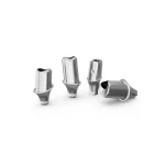

With the acceleration of global population aging and the rising awareness of oral health, dental implants have become the preferred solution for restoring missing teeth. Statistics show that the number of new dental implants worldwide has exceeded ten million cases annually, while the Chinese market continues to maintain a growth rate of over 20% per year. This technological advancement has significantly improved patients’ chewing function and quality of life. However, it has also raised an increasingly prominent clinical issue: when patients need to undergo magnetic resonance imaging (MRI), will the metallic components of the implants interact with the strong magnetic field, potentially interfering with image quality or even threatening examination safety?
This article will systematically review the mechanisms of interaction between various implant components and MRI, drawing on guidelines from international authoritative organizations and data from cutting-edge research. It will analyze the extent to which different materials and scanning sites affect imaging quality and propose clinical management recommendations based on risk stratification. By dispelling the misconception that “metal automatically equals contraindication,” the aim is to establish a scientific and safe decision-making framework for both clinicians and patients in balancing implant treatment planning and medical imaging needs.
Material Properties of Dental Implants
As the core device replacing natural tooth roots in modern dentistry, the choice of material for dental implants directly determines their biocompatibility, mechanical stability, and compatibility with medical imaging. Current mainstream implant materials can be categorized into two main systems: metal-based materials (represented by pure titanium/titanium alloys) and non-metallic ceramic materials (centered on zirconia). These two systems exhibit significant differences in physical properties, MRI response, and clinical application scenarios.
1. Metal-Based Implants
Pure titanium (Ti) and titanium alloys (such as Ti-6Al-4V) are the most widely used implant materials in clinical practice. Their MRI compatibility stems from their unique paramagnetic properties:
- Extremely low magnetic susceptibility: The volumetric magnetic susceptibility of titanium is +182 × 10⁻⁶ (SI units), which is only 1/1000 that of ferromagnetic materials. The induced torque in 1.5T/3.0T MRI magnetic fields is negligible, posing virtually no risk of magnetic displacement.
- Controlled thermal effects: The local temperature rise induced by RF pulses does not exceed 0.5°C, far below the 1°C safety threshold set by the International Electrotechnical Commission (IEC), ensuring no risk of tissue burns.
- Limited artifact size: In 3.0T MRI, pure titanium generates metal artifacts with a diameter typically <5 mm, exerting minimal impact on the visualization of adjacent anatomical structures, especially suitable for implants in the posterior region.
Based on these properties, titanium implants are classified under international standards (ASTM F2503) as “MRI Conditional”—meaning they are safe under specific conditions (such as magnetic field strength and scanning sequences). It should be noted that alloying elements such as aluminum and vanadium may slightly increase magnetic susceptibility, but clinical studies have confirmed that they remain within safety standards.
2. Non-Metallic Ceramic Implants: Complete Magnetic Inertness of Zirconia
Zirconia (ZrO₂), a representative of all-ceramic restorative materials, has recently gained prominence in the implant field, offering absolute advantages in MRI compatibility:
- Zero magnetic response: As a non-metallic ceramic oxide, zirconia’s magnetic susceptibility is close to zero. It generates no induced currents or displacement effects in MRI magnetic fields, completely eliminating magnetic attraction risks.
- No imaging artifacts: Ceramic materials do not reflect RF pulses, producing no metal artifacts in imaging. This makes zirconia especially suitable for aesthetic restorations in the anterior region and for head and neck MRI (e.g., temporomandibular joint, salivary gland examinations).
- Outstanding biocompatibility: The oxide layer on zirconia surfaces inhibits bacterial adhesion. Its elastic modulus (210 GPa) is closer to that of natural dentin, helping to reduce the risk of bone resorption.
Despite zirconia’s perfect MRI compatibility, it presents challenges such as high manufacturing difficulty and intrinsic brittleness. At present, it is mostly used for single-tooth restorations or short-span bridges, with long-term clinical data still under accumulation.
3. MRI Compatibility Labeling and Material-Based Clinical Selection
The MRI compatibility of implants made from different materials should be judged comprehensively based on manufacturer labeling and clinical context:
- Titanium implants:
- MRI Safe: Some manufacturers optimize surface treatments (e.g., SLA hydrophilic coatings) to further reduce susceptibility, labeling their products as “MRI Safe”, usable under any field strength.
- MRI Conditional: Most titanium implants fall into this category, requiring conditions such as:
- Magnetic field strength ≤ 3.0T
- Gradient field strength ≤ 40 T/m
- Specific scan sequences (e.g., avoiding gradient echo sequences)
- Typical labeling example: “Safe up to 3T when placed in the maxilla/mandible.”
- Zirconia implants:
- As inherently non-magnetic materials, manufacturers usually label them directly as “MRI Safe”, with no conditional restrictions.
4. Clinical Relevance of Material Properties
- Posterior functional restorations: Titanium implants are preferred for their high mechanical strength (fracture resistance > 900 MPa), which can withstand chewing loads. MRI artifacts in these regions typically have little diagnostic impact.
- Anterior aesthetic restorations / Head & neck MRI requirements: Zirconia implants are the better choice, as their artifact-free property ensures clear imaging of facial nerves, blood vessels, and tumor margins.
- Special patient populations: For patients requiring frequent MRI scans (e.g., oncology patients), it is advisable to consult with radiologists before surgery and prioritize implant systems explicitly labeled as “MRI Safe”.
MRI and Metal Implants: The Triangular Relationship of Magnetic Fields, Metals, and Imaging Safety
Magnetic resonance imaging (MRI), as a core imaging technology in modern medicine, operates on principles that interact in complex ways with metallic implants, forming a physical-biomedical system. Understanding this system requires analysis from three perspectives: the fundamentals of MRI imaging, the electromagnetic responses of metals, and the resulting clinical risks.
1. Physical Basis of MRI Imaging: Synergy of Strong Magnetic Fields and Radiofrequency Pulses
The core imaging principle of MRI is based on the nuclear magnetic resonance phenomenon, which involves three key stages:
- Static magnetic field establishment:
Superconducting magnets generate strong static magnetic fields ranging from 1.5T to 7T (the Earth’s magnetic field is about 50 μT, meaning MRI fields are 30,000 to 140,000 times stronger). These fields align the magnetic moments of hydrogen protons (primarily found in water and fat) along the field direction (Z-axis). - Radiofrequency (RF) pulse excitation:
RF pulses at specific frequencies are emitted perpendicular to the static magnetic field, causing hydrogen protons to absorb energy, precess, and deviate from the Z-axis into the transverse plane (XY-plane), thereby forming a macroscopic transverse magnetization vector. - Signal reception and image reconstruction:
When RF pulses stop, hydrogen protons release energy via free induction decay (FID), generating weak alternating electromagnetic signals (MR signals). Receiving coils capture these signals, which are then transformed into anatomical images through Fourier reconstruction.
Key point: MRI image quality relies heavily on the homogeneity of the static magnetic field and the precision of RF pulse control. Any external magnetic substances or electromagnetic interference can disrupt this balance, causing image distortion or equipment malfunction.
2. Interactions Between Metal Implants and MRI: Three Major Risk Mechanisms
The risks associated with metal implants in MRI environments stem from their electromagnetic and thermodynamic behaviors, manifesting in three main effects:
(1) Magnetic attraction and displacement risks: The dominance of magnetic forces on ferromagnetic materials
- Ferromagnetic materials (e.g., iron, nickel, cobalt, and their alloys) have high magnetic permeability (μ ≫ μ₀). In strong magnetic fields, they are rapidly magnetized and subjected to forces proportional to the field gradient:
F=χV∇B2/2μ0F = χV∇B² / 2μ₀F=χV∇B2/2μ0
where χ = magnetic susceptibility, V = volume, B = magnetic flux density. - Clinical consequences:
- Small implants (e.g., surgical needles, fragments) may be rapidly pulled toward the magnet, causing tissue tearing or equipment damage.
- Large implants (e.g., joint prostheses, vascular stents) are less likely to shift entirely but may undergo micromovements, leading to wear, material fatigue, or metal ion release.
- Example case: In 2001, the U.S. FDA reported that a ferromagnetic oxygen tank was pulled into a 1.5T MRI bore, causing severe cranial injury to a patient.
(2) Image artifact interference: Dual disturbances from metals on RF pulses and static fields
- Static field inhomogeneity:
Metal implants, especially magnetic ones, distort local magnetic fields, causing inconsistent precession frequencies of hydrogen protons. This results in striped black-and-white artifacts in images (e.g., ghosting artifacts, chemical shift artifacts).- The artifact size is proportional to the metal’s volume, shape, and magnetic field strength. In 3.0T MRI, the artifact diameter can reach 5–10 times the implant’s size.
- RF pulse reflection and absorption:
Metals’ high electrical conductivity (σ) makes them reflectors of RF pulses, causing signal void artifacts. Simultaneously, absorbed RF energy is converted into heat (discussed below). - Clinical impacts:
- In head and neck MRI, metallic implants may completely obscure nearby structures (e.g., mandibular nerve canal, temporomandibular joint), leading to diagnostic information loss.
- In spinal MRI, metallic fixation devices may mask disc herniations or tumors.
(3) Metal heating effects: RF-induced Joule heating and tissue damage
- Mechanism:
Alternating RF magnetic fields (at frequencies matching the Larmor frequency) induce eddy currents in metals. According to Joule’s law (Q = I²Rt), these currents generate heat as they flow through the metal’s resistance (R). - Intensity factors:
Heating correlates positively with the metal’s electrical conductivity (σ), RF pulse power (SAR value), and exposure duration. - Heat transfer and tissue injury:
Poor heat dissipation at the metal–tissue interface can form local hotspots. A temperature rise above 43°C may cause protein denaturation and cell death. Sensitive tissues (e.g., spinal cord, optic nerve) can be damaged by increases as small as 1–2°C. - Safety standards:
The International Electrotechnical Commission (IEC) stipulates that tissue heating around implants during MRI must not exceed 1°C (at SAR ≤ 2 W/kg). Titanium alloys, with relatively low conductivity (σ ≈ 2.5 × 10⁶ S/m), pose significantly lower heating risks compared to stainless steel (σ ≈ 1.4 × 10⁶ S/m).
The Practical Impact of Dental Implants on MRI Examinations
As a commonly used device for oral rehabilitation, the compatibility of dental implants with MRI examinations directly affects both patient management and imaging quality. Based on material properties and clinical studies, the impact of implants on MRI can be systematically analyzed from two dimensions—safety and image quality—to provide scientific evidence for clinical decision-making.
I. Safety Assessment
The core safety risks of MRI examinations arise from the mechanical effects of strong magnetic fields on implants and the thermal effects of radiofrequency (RF) pulses. Titanium and zirconia implants, due to their unique material properties, demonstrate a high level of safety in MRI environments.
1. Magnetic Attraction and Displacement Risk: Zero or Negligible
- Titanium Implants:
Pure titanium (Ti) and titanium alloys (e.g., Ti-6Al-4V) are paramagnetic materials. Their magnetic susceptibility (χ ≈ +182 × 10⁻⁶) is only 1/1000 that of ferromagnetic materials. The magnetization force generated in 1.5T/3.0T MRI is extremely low.- Clinical study: A 2018 Journal of Oral Implantology study showed that in 3.0T MRI, the maximum displacement of titanium implants was <0.1 mm, far below the clinical safety threshold (0.5 mm), with no reports of dislodgment or displacement.
- Zirconia Implants:
Zirconia (ZrO₂) is a non-metallic ceramic with near-zero magnetic susceptibility, showing no magnetic response, and therefore no attraction or displacement effects in magnetic fields.- Typical case: A 2021 Clinical Oral Implants Research report confirmed that zirconia implants remained stable even under 7T MRI (an ultra-high-field research system), verifying their absolute safety.
2. RF-Induced Heating Effects
- Titanium Implants:
Titanium has relatively low electrical conductivity (σ ≈ 2.5 × 10⁶ S/m), limiting the strength of eddy currents induced by RF pulses. In 3.0T MRI, implant surface temperature typically increases by <0.3°C, and surrounding tissue by <0.5°C—well below the IEC safety threshold of 1°C.- Influencing factors: Temperature rise is related to implant surface area, RF power (SAR value), and scan duration. Short sequences (e.g., T1WI) pose much lower heating risks than long sequences (e.g., DWI).
- Zirconia Implants:
As an insulator (σ ≈ 10⁻¹² S/m), zirconia generates no eddy currents, and RF-induced heating is negligible, with tissue temperature rise close to baseline.
3. Clinical Consensus
According to international standards (ASTM F2503), both titanium and zirconia implants are classified as “MRI Conditional.” However, due to the absence of magnetic response, zirconia’s actual safety profile is closer to the “MRI Safe” standard.
II. Impact on Image Quality
The main mechanisms by which implants affect MRI images are static field inhomogeneity and RF pulse reflection, with manifestations closely related to implant location, material, and scan sequence.
1. Maxillofacial MRI: Local Artifacts Are Significant but Diagnostic Value Is Retained
- Artifact Mechanisms:
- Titanium implants: Paramagnetism distorts the local magnetic field, producing black-and-white band-like artifacts (susceptibility artifacts), typically 3–5 times the implant diameter (3.0T MRI).
- Zirconia implants: With no magnetic response, artifacts stem only from RF reflections, appearing as signal voids <2 mm—significantly smaller than titanium artifacts.
- Clinical Impact:
- Single-tooth implants: Artifacts may obscure adjacent tooth roots or alveolar bone, but adjusting the scan plane (e.g., oblique sagittal) can partially avoid them.
- Multiple-tooth bridges: Overlapping artifacts may affect the assessment of mandibular continuity, but not the evaluation of implant stability (e.g., osseointegration).
- Typical case: A 2020 Dentomaxillofacial Radiology study found titanium implant artifacts in 3.0T MRI covered ~15 mm², but metal artifact reduction sequences (MARS) reduced this to ~5 mm².
2. MRI of Regions Remote from the Jaws
- Brain MRI:
Since implants are in the jaws and >5 cm away from brain tissue, magnetic field gradient decay reduces to <1% of baseline, with no artifact interference.- Clinical validation: A 2019 American Journal of Neuroradiology study of 100 implant patients found no statistically significant difference (p > 0.05) in brain MRI image quality scores compared with non-implant controls.
- Spinal MRI:
In cervical MRI, when implants are >3 cm from the spinal cord, artifacts do not affect intradural structure evaluation. Lumbar MRI is completely unaffected.
3. Sequence Optimization: Clinical Application of Artifact Suppression
- Metal Artifact Reduction Sequence (MARS):
By increasing bandwidth, adjusting flip angles, and using presaturation pulses, MARS can reduce titanium implant artifacts by 30%–50%, especially useful in evaluating jaw tumors or inflammation. - High-resolution T2-weighted imaging (T2WI):
For zirconia implants, T2WI can clearly visualize surrounding soft tissues (e.g., gingiva, mucosa) with minimal artifact interference.
III. Clinical Decision-Making Recommendations: Individualized Choices Based on Risk–Benefit Analysis
- Emergency MRI (e.g., stroke, spinal cord injury):
No need to delay scans due to implants. Titanium and zirconia implants do not affect diagnoses of regions distant from the jaws. - Maxillofacial tumor or inflammation assessment:
Prefer zirconia implants or MARS sequences to minimize artifacts obscuring lesion margins. - Long-term follow-up patients:
Zirconia implants, due to their lack of magnetic response, are better suited for patients requiring frequent head and neck MRI scans (e.g., post-radiotherapy monitoring for nasopharyngeal carcinoma).
Conclusion
Both titanium and zirconia implants demonstrate a high level of safety during MRI examinations. Their impact on image quality is largely confined to the maxillofacial region and can be significantly reduced through sequence optimization. Clinical decision-making should comprehensively balance implant material, scanning site, and diagnostic requirements, ensuring safety while maximizing imaging diagnostic value. With the maturation of zirconia implant technology and the wider adoption of MARS sequences, the practical limitations of implants in MRI examinations are gradually diminishing, opening broader opportunities for precise diagnosis and treatment of oral and maxillofacial diseases.